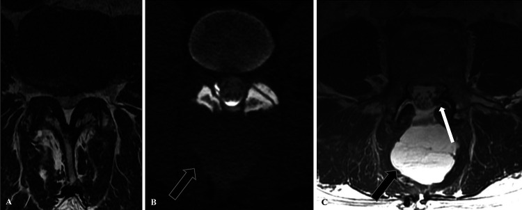
FIG. 1.
Case 1. A: Preoperative axial MRI at the L3–4 level demonstrated severe central and bilateral lateral recess stenosis. B: Postoperative CT myelogram with evidence of epidural contrast leakage into a large dorsal paraspinous fluid collection (arrow), suspicious for CSF leakage. C: Postoperative T2 FIESTA MRI highlights thickening and displacement of the lumbosacral nerve roots within the diastatic left L3–4 facet joint (white arrow), along with a large subfascial fluid collection (black arrow).

