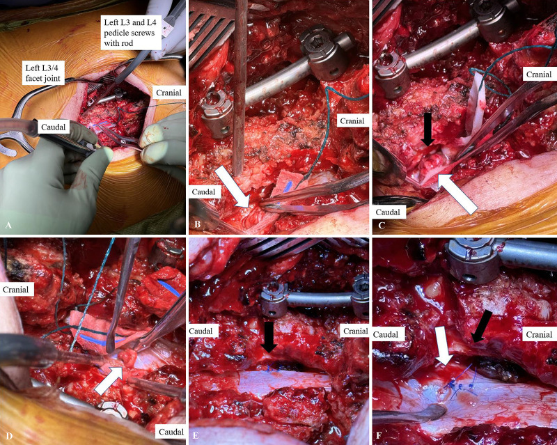
FIG. 2.
Case 1. A: The thecal sac has been exposed. Note that the left L4 nerve root herniation is not immediately visible, because it remains entrapped in the L3–4 facet joint. On the patient’s left side, pedicle screws have been placed at L3 and L4, accompanied by a rod. B: After rod distraction, the left L4 nerve root herniation (arrow) is now visible and can be retrieved from the left L3–4 facet joint. C: Note the proximity of the herniated left L4 nerve root (white arrow) to the left L3–4 facet joint line (black arrow). D: Viewed from the patient’s left, one can appreciate the herniated left L4 nerve root (arrow), with a swollen and erythematous appearance. Also in this image are the right L3 and L4 pedicle screws, without a rod connecting at this point in the procedure, because no distraction was necessary on this side. E: View from the patient’s right after the left L4 nerve root has been reduced into the thecal sac and the durotomy repaired. Note the proximity of the left L4 nerve root shoulder (black arrow) to the left L3–4 facet joint in which it was previously entrapped. F: Note the dural repair at the medial-most aspect of the nerve root shoulder (white arrow) and the wide medial facetectomy performed to access the dura for repair and ensure adequate decompression of the neural elements.

