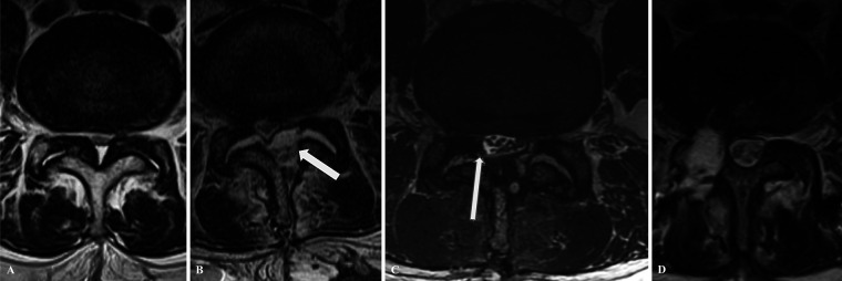
FIG. 3.
Case 2. A: Preoperative axial MRI at the L4–5 level showing severe central and lateral recess stenosis. B: Postoperative axial T2-weighted MRI demonstrates an over-the-top decompression extending from the left hemilamina to the right lateral recess, with a compressive fluid collection (arrow). C: MRI with the T2 FIESTA sequence highlights the right-sided lumbosacral nerve rootlets entrapped in the diastatic right L4–5 facet joint (arrow). D: Six-month postoperative axial T2-weightde MRI demonstrates dural repair with evidence of the maintained reduction of the previously herniated nerve roots. Also evident is a complete right-sided facetectomy.

