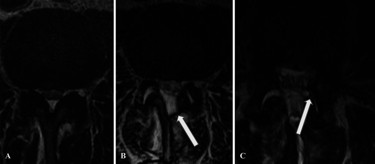
FIG. 4.
Case 3. A: Preoperative axial MRI at the L3–4 level demonstrates severe central and bilateral lateral recess stenosis. B: Postoperative MRI demonstrates a postoperative fluid collection (arrow) at the surgical site, along with diastasis of the bilateral L3–4 facet joints. C: Axial T2-weighted MRI after the evacuation surgery demonstrates eventration of the thecal sac contents into the left L3–4 facet joint (arrow).

