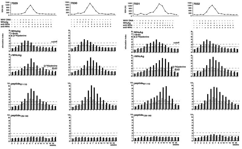
FIG. 1.
Humoral and cellular immune responses of woodchucks NW7029, NW7030, NW7031, and NW7032 during an acute, self-limited WHV infection. T-cell responses to WHsAg, rWHcAg, peptide 97–110, and peptide 129–140 were monitored during WHV infection with 105 woodchuck ID50. WHsAg, anti-WHs, and anti- WHc in the sera were detected by ELISA. SDH levels were assessed by a commercial enzyme assay. Viremia was detected by DNA dot blot (boldface +) and PCR (lightface +). T-cell responses (5 × 10⁴ PBMC) after stimulation with 2 μg of WHsAg per ml or 1 μg of rWHcAg or peptides per ml were analyzed weekly after infection by BrdU and [2-3H]adenine incorporation. Results are presented as mean SI of triplicate determinations. The mean values (optical density at 450 nm) for the controls were 0.22 ± 0.08. The mean cpm for the controls was 3,213 ± 1,722.

