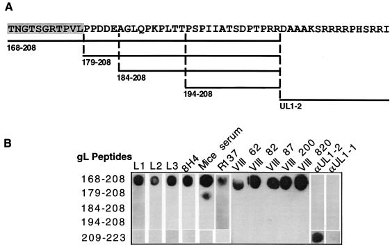
FIG. 6.
Mapping of anti-gL antibody epitopes with synthetic peptides. (A) Diagram depicting the sequences of the set of overlapping synthetic peptides mimicking the gL sequence. The location of each peptide within the gL sequence is indicated. (B) Dot blot analysis of anti-gL antibodies with the peptides. Two microliters of each peptide (4 μg/dot) was spotted onto nitrocellulose membrane strips. After blocking, antibodies were added to each strip, and the reactivity was detected by ECL with goat anti-mouse peroxidase or goat anti-rabbit peroxidase.

