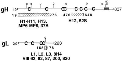
FIG. 7.
Positions of the epitopes for MAbs to gH and gL mapped in this study. Schematic figures depict the linear amino acid sequences of gH and gL. The hatched bars depict the locations of the epitopes of anti-gH and anti-gL antibodies. The position of MAb 52S is according to the amino acid change (residue 536) of two MAR mutants selected by 52S (13).

