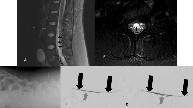
Figure 2.
Lumbar imaging confirming persistent ventral cerebrospinal fluid leak. (A and B) Midline sagittal (A) and axial (B, lumbar L3 level) T2-weighted fat suppressed MRIs. Black arrows show ventral dura (A and B). White arrow shows ventral epidural fluid collection (B). (C–E) Prone digital subtraction myelogram: (C) lateral image of lumbar spine with patient prone on cath lab table showing needle placement and demonstrating site of contrast injection. (D) Early postcontrast injection images show pooling of unmixed intrathecal contrast along the ventral dura (black arrows) and ventral extravasation of contrast (gray arrow) at L3. (E) Seconds-delayed postcontrast injection image shows further spread of ventral epidural contrast extending from the ventral dural defect (gray arrow).

