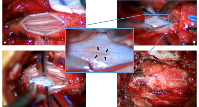
Figure 3.
Intraoperative imaging. (A) Dorsal dura has been opened in the midline and tacked open with 6-0 prolene sutures after an L3 laminectomy was performed. Cauda equina is visualized. (B) After careful inspection of visualizable ventral dura within the extent of our exposure, nerve roots have been retracted laterally and a defect is identified in the ventral dura (blue rectangle). (C) Magnified view of the ventral elliptical dural defect (black arrows show edges). (D) A 6-0 Gore-Tex suture has been used to primarily repair the ventral dural defect. (E) The dorsal dura has been repaired primarily with 6-0 Gore-Tex suture.

