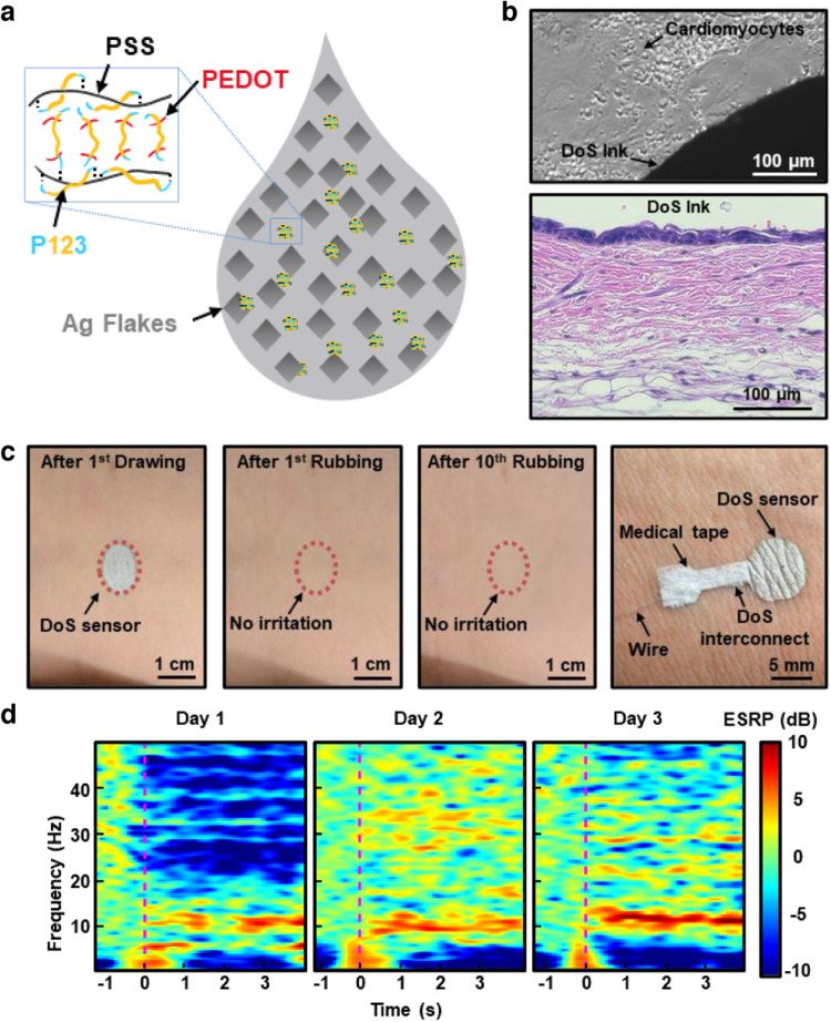
Fig. 2. Biocompatible DoS conductive ink.
a Schematic of one possible arrangement of PEDOT:PSS, Ag flakes, and P123 in the DoS conductive ink. b Microscope image of cardiomyocytes co-cultured with DoS ink (top) and skin histology of mice skin exposed to DoS conductive ink for 3 days (bottom). c Drawing and erasing for DoS sensors multiple times on the skin (left 3 frames). One approach to wire the DoS sensor is shown in the right frame. d Recording of EEG signals during mental relaxation recorded on 3 consecutive days. Reprinted with permission from Yu et al.11. Copyright 2022 WILEY-VCH.

