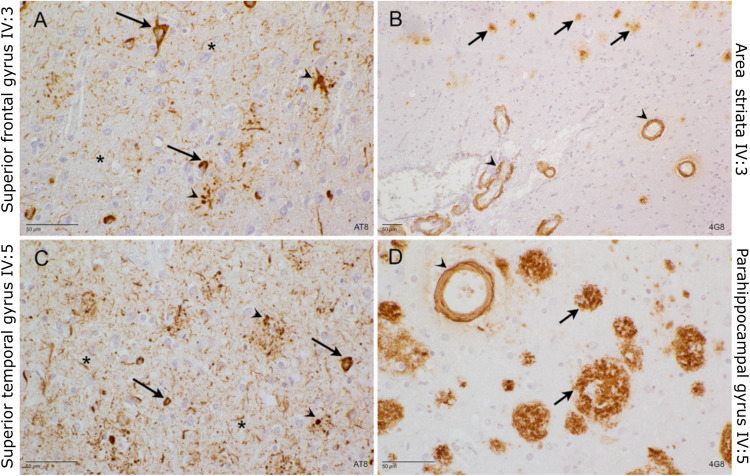
Fig. 2. Neuropathological examination of brain tissue of IV:3 and VI:5.
A Superior frontal gyrus of IV:3: AT8 stain. Many neurofibrillary tangles (arrow), as well as dystrophic neurites (arrowhead) and neuritic threads (asterisk) are detected. B Area striata of IV3: 4G8 stain. Small magnification to show the abundance of cerebral amyloid angiopathy (CAA, arrowhead) and senile plaques (arrow). C Superior temporal gyrus of IV:5 AT8 stain. Many neuritic threads (asterisk) and dystrophic neurites (arrowhead) affect the temporal cortex. Many neurons contain neurofibrillary tangles (arrow). D Parahippocampal gyrus of IV:5: 4G8 stain. Many large senile plaques (arrow), again in the presence of cerebral amyloid angiopathy (arrowhead).

