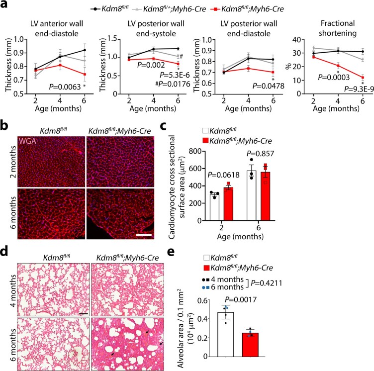
Extended Data Fig. 2. Kdm8 mutants develop dilated cardiomyopathy with no signs of concentric hypertrophy.
a, Echocardiogram analysis of left ventricle (LV) anterior wall thickness at end-diastole, LV posterior wall thickness at end-systole, LV posterior wall thickness at end-diastole, and LV fractional shortening in control (Kdm8fl/fl), heterozygous (Kdm8fl/+;Myh6-Cre), and mutant (Kdm8fl/fl;Myh6-Cre) mice at 2, 4 and 6 months of age. Error bars denote the mean + /- s.e.m. Data was analyzed by 2-way ANOVA with Tuckey’s multiple comparison correction. * denotes comparisons between control and mutants, # between control and heterozygous hearts. n = 6 - 11 mice per group. b, Wheat germ agglutinin staining (WGA) on cross sections of the hearts of 2 and 6-month-old control and mutant mice. Scale bar, 100 μm. c, Cell surface area quantified from sections stained with WGA and analyzed by two-tailed Student’s t-test. Error bars denote the mean + /- s.e.m. n = 3 hearts per group. d, Lung sections of control and Kdm8 mutants at 4 and 6 months of age stained with H&E. Arrows point to areas of accumulated alveolar fluid. Scale bar = 100 μm. e, Alveolar area per 0.1 mm2 in lung sections Error bars denote the mean + /- s.d. Data was analyzed by two-way ANOVA with Sidak correction. n = 3 4-month-old and 2 6-month-old lungs of controls, and 2 4-month-old and 2 6-month-old lungs of mutant mice.

