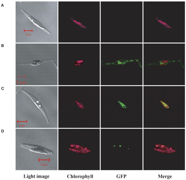
Figure 2.
Fluorescent microscope images of wild-type cells and transformants of P. tricornutum. Wild-type cell (A); transformants with construct e (B); with construct a (C); and with construct b (D). Light images, chlorophyll autofluorescence, GFP fluorescence, and merged images of chlorophyll and GFP fluorescence are presented, respectively, from the left column. Images were taken with a laser-scanning confocal microscope under conditions described in the text. Scale bars, 5 μm. Cells were grown in air-level (0.037%) CO2.

