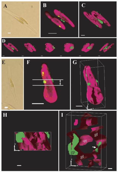
Figure 4.
Analyses of a three-dimensional model of inner chloroplast structures in two independent pre138-mptca1-egfp transformants of P. tricornutum by sectioning fluorescent microscopy. A light image (A); a merged image of chlorophyll and GFP fluorescence of the corresponding portion to A (B); a three-dimensional model of B (C); X-axial rotation of the three-dimensional model (D); a light image (E); a merged fluorescence image of chlorophyll and GFP fluorescence of the corresponding portion to E (F); a three-dimensional model of F (G); a surface image of the cross-sectioned portion indicated in F (H); and a cross-section image that is rotated on the x axis about 90° from H (I). Magenta parts indicate chlorophyll fluorescence and green parts indicate GFP fluorescence. Images in D were taken at every 30° rotation. The cross-sectioned portion of 1.0-μm thickness is indicated in F. x, y, and z axes indicate the topological relationship among G, H, and I. gl, Girdle lamellae; ct, central thylakoid stacks; gp, GFP particle. Scale bars, 2 μm in A, B, E, and F; 1 μm in C, D, and G; 0.2 μm in H and I. Cells were transformed with the construct b in Figure 1B and grown in air-level CO2.

