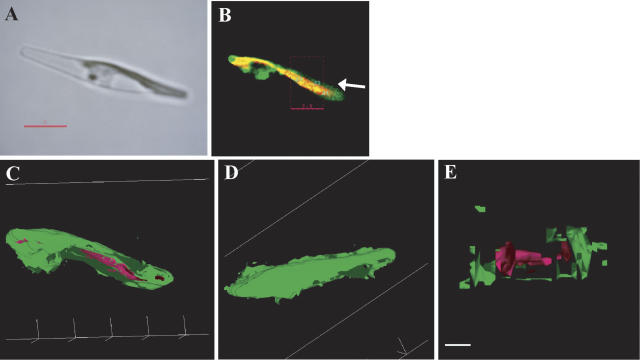
Figure 6.
Analyses of a three-dimensional model of the chloroplast in a pre1-66-mptca1-egfp transformant. A light image (A); a merged image of chlorophyll and GFP fluorescence of the corresponding portion to A (B); a three-dimensional model viewed from the bottom of B (C); viewed from the top of B (D); and a cross-section image of the model (E). Magenta parts indicate chlorophyll fluorescence and green parts indicate GFP fluorescence. The cross-sectioned portion of the chloroplast is indicated by dashed box in B. The thickness of the section was 3.5 μm. Scale bar in A, 5 μm; scale bar in (E), 2 μm. Cells were transformed with construct c in Figure 1B and grown in air-level CO2.

