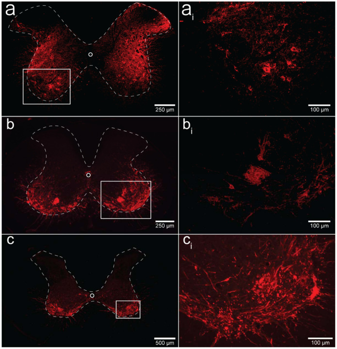
Figure 6. Histological assessment of mCherry expression in the C4/C5 spinal segments.
Representative photomicrographs of mid-cervical spinal sections from a wild-type mouse (a-ai), a ChAT-Cre mouse (b-bi), and a ChAT-Cre rat (c-ci). Wild-type mice (a-ai) showed a nonspecific pattern of expression throughout the mid-cervical grey matter. ChAT-Cre mice and rats (b-ci) showed expression limited to neurons in the ventral horns. Red color indicates positive and mCherry fluorescence. Dashed white line indicates the approximate white-gray matter demarcation.

