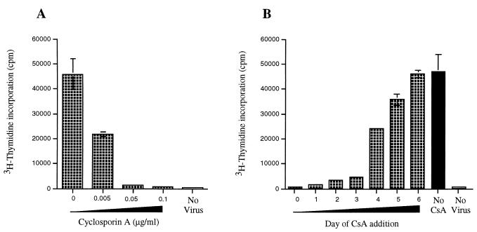
FIG. 4.
(A) PBMCs were infected with PBj6.6 virus, or mock-infected, and incubated in the presence of the indicated concentrations of cyclosporin A. (B) PBMCs were infected with PBj6.6 virus, or mock-infected, and CsA (0.1 μg/ml) was then added to the cultures at the indicated times after virus infection (days); proliferation was measured 7 days after initial exposure of the cells to PBj6.6 virus. Values shown represent the means from a single experiment that was performed in triplicate; the standard errors of mean values are marked by bars. The results shown are representative of two experiments that yielded similar results; in addition, the experiment whose results are shown in Panel A was repeated on three occasions, with similar results, with a different molecular clone of SIVsmmPBj14—PBj 1.9 (7, 12).

