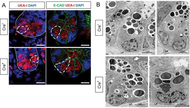
Figure 4. Glial depletion triggers morphological changes in Paneth cells.
A) Representative images of UEA-I staining of Paneth cell granules in the small intestine of Cre− and Cre+ mice (observed in at least 3 mice per genotype). Scale bar = 10μm.
B) Representative transmission electron microscopy images of Paneth cells (n = 2 mice per genotype from independent cohorts). Paneth cells in Cre+ mice are globular, exhibit loss of polarity, and have heterogeneous granules (PG). L, Lumen of the intestinal crypts; LP, lamina propria. Scale bar = 3μm.

