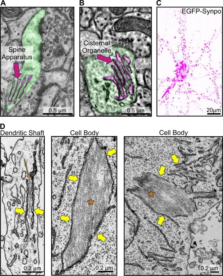
Fig. 1. Synaptopodin mediated recruitment of actin filaments to the ER in neurons.
A. Transmission electron microscopy (TEM) image of a pseudocolored dendritic spine (green) with spine apparatus (magenta). B. Serial block-face scanning electron microscopy (SBF-SEM) image of a pseudocolored cisternal organelle (magenta) at an axonal initial segment (green) (image from microns-explorer.org)[38]. C–D. Fluorescent image (C), and TEM (D) of a cultured mouse hippocampal neuron infected with AAV2/9 encoding EGFP-synaptopodin. Note in C large accumulations of synaptopodin in the central region of the cells in addition to the punctate fluorescence in dendrites. Many of smaller puncta along dendrites correspond to the expected accumulations of synaptopodin in the spine apparatus of dendritic spines. Yellow arrows in D point to the ER and one orange arrow and asterisks point to actin bundles.

