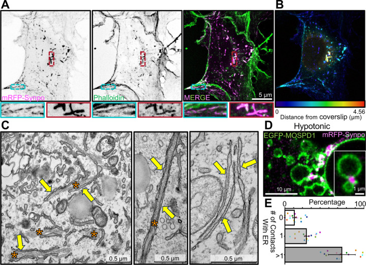
Fig. 2. Localization of exogenous synaptopodin in COS-7 cells both on stress fibers and on actin-rich structures associated with ER.
A. COS-7 cells expressing mRFP-synaptopodin displaying strong overlap of the mRFP fluorescence with phalloidin staining. High magnification views of a stress fiber (blue) and a non-stress fiber linear assembly (red) are shown at the bottom. B. mRFP-synaptopodin signal from A is shown with color-coding based on the distance from the coverslip. Stress fibers are closer to coverslip while non-stress fiber actin assemblies are present anywhere within the cell. C. TEM of a cell expressing mRFP-synaptopodin showing actin bundles (orange asterisks) sandwiched between ER sheets (yellow arrows). D. COS-7 cells expressing mRFP-synaptopodin (magenta) and the ER protein EGFP-MOSPD1 (green) were exposed to hypotonic conditions. The accumulation of synaptopodin at the interface between ER elements reveals its direct or indirect association with the ER membrane. E. Percentage of mRFP-synaptopodin puncta with or without association with ER membrane in COS-7 cells exposed to hypotonic conditions is shown. Data from different cells are shown as a dot with different color.

