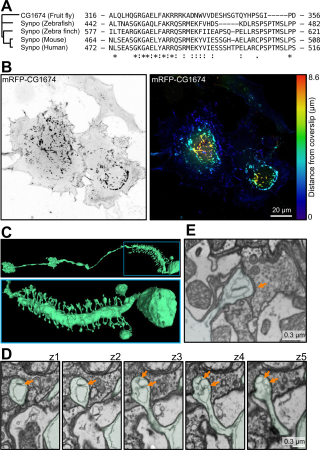
Fig. 4. Presence of synaptopodin and spine apparatus in Drosophila melanogaster.
A. Residues 464–508 of mouse synaptopodin (calsarcin domain) are highly conserved among synaptopodin orthologues in vertebrates and D. melanogaster orthologue, CG1674. B. COS-7 cell expressing the fluorescently tagged D. melanogaster synaptopodin orthologue. A prominent accumulation of CG1674 inclusions, similar to those observed in cells expressing fluorescently tagged mouse synaptopodin (See Fig. 2 and 3C), is visible. The CG1674 signal is shown in gray scale (left), and color-coded based on distance from coverslip (right). C. D. melanogaster ‘s LPTC neuron reconstructed from 3D SBF-SEM images (from FlyWire [46]) showing spines along its major process. Low and high magnification views are shown at the top and bottom, respectively. D. SBF-SEM sequential optical sections from a spine of the neurons shown in C, revealing ER elements closely apposed via an intervening density (orange arrows) in the spine head. This structure is reminiscent of a spine apparatus with dilated ER cisterns, possibly due to preparation artifacts. E. Another example of a dendritic spine of D. melanogaster containing a structure reminiscent of a spine apparatus, but with a dilated ER.

