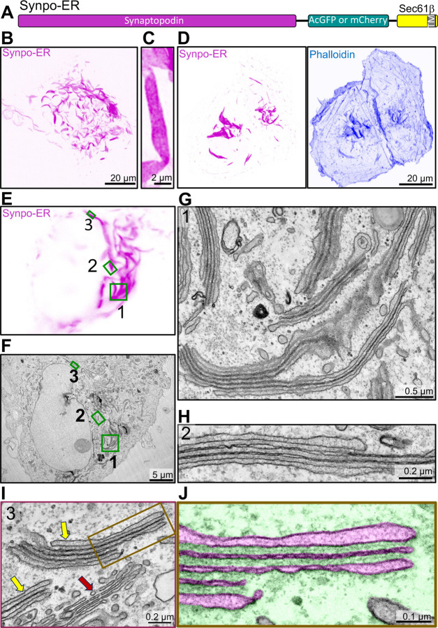
Fig. 5. Generation of spine apparatus-like structures (SALs) in COS-7 cells.
A. Diagram of the synaptopodin construct anchored to the ER (Synpo-ER) by its fusion to Sec61β, which is embedded in the ER by a C-terminal transmembrane region. B. Confocal image of a COS-7 cell expressing Synpo-ER. C. High magnification view of a SAL as imaged by an AiryScan confocal microscope. D. Presence of F-actin in SALs as indicated by phalloidin staining. E and F. Light microscope image (E) of a COS-7 cell expressing synaptopodin with its corresponding EM image (F). High magnification EM images of the regions 1 – 3 framed by green rectangles in fields E and F are shown in G–I, respectively. Moreover, a portion of the SAL in I is shown at higher magnification in J with the ER lumen pseudocolored in magenta and the cytosolic space in green. SALs are represented by ER stacks with morphological features similar to those of the spine apparatus and of the cisternal organelle. Red and yellow arrows in I point to the Golgi complex and to two SALs, respectively.

