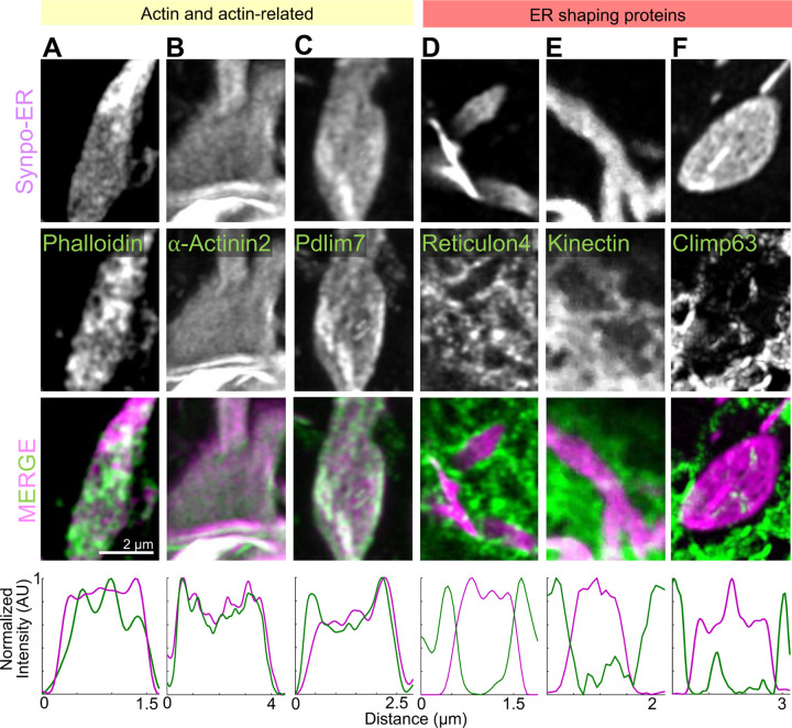
Fig. 6. Presence of actin and actin-related proteins in SALs. A–F.
High magnification AiryScan images of individual SALs showing the localization of Synpo-ER and of the fluorescently tagged proteins indicated. Line scan plots are shown in the bottom. Reticulon 4-GFP is enriched at the edges of SALs and excluded from their flat portions. GFP-Kinetctin and GFP-Climp63 are excluded from SALs.

