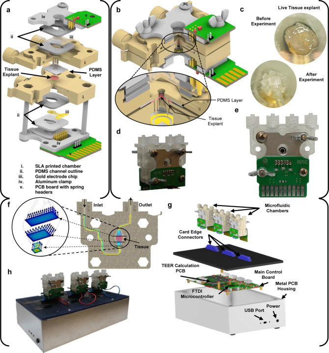
Figure 1: Major components of the microphysiological system.
Including the microfluidic chamber (a-f) and the system-level housing (g-h). a) Expanded view of a full chamber, with all components labelled. b) Closed chamber with a closeup view of the tissue and PDMS clamped between two chamber halves. c) Tissue explant before and after the experiment. d) and e) The actual manufactured chamber assembled (front and back, respectively). f) Flow simulation through the chamber’s microfluidics. g) Expanded view of the microphysiological system. h) The actual manufactured and fully assembled system with three chambers connected the system.

