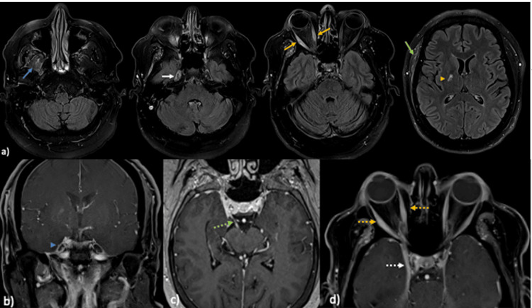
Figure 3. Brain and orbit MRI with contrast .
(a) Axial magnetic resonance imaging (MRI) T2 fluid-attenuated inversion recovery (FLAIR) images of the brain and orbits show asymmetric thickening and hyperintensity of the right-sided masticator muscles (blue arrow), the right ganglion of trigeminal nerve (white arrow), and all extraocular muscles in the right orbit (orange arrows) and right temporalis muscle (green arrow). There is an associated subacute lacunar infarct in the posterior limb or the right internal capsule (orange arrowhead). (b)-(d) Contrast MRI of the brain and orbit coronal T1 fat saturated with contrast (b), axial T1 fat saturated with contrast (c) and axial T1 fat saturated with contrast (d) showed abnormal asymmetric enhancement of the right ganglion of the trigeminal nerve (blue arrow head) and the right oculomotor nerve (green arrow), asymmetric thickening and enhancement of the right extraocular muscles (dotted orange arrows) and the right cavernous sinus (white dotted arrow).

