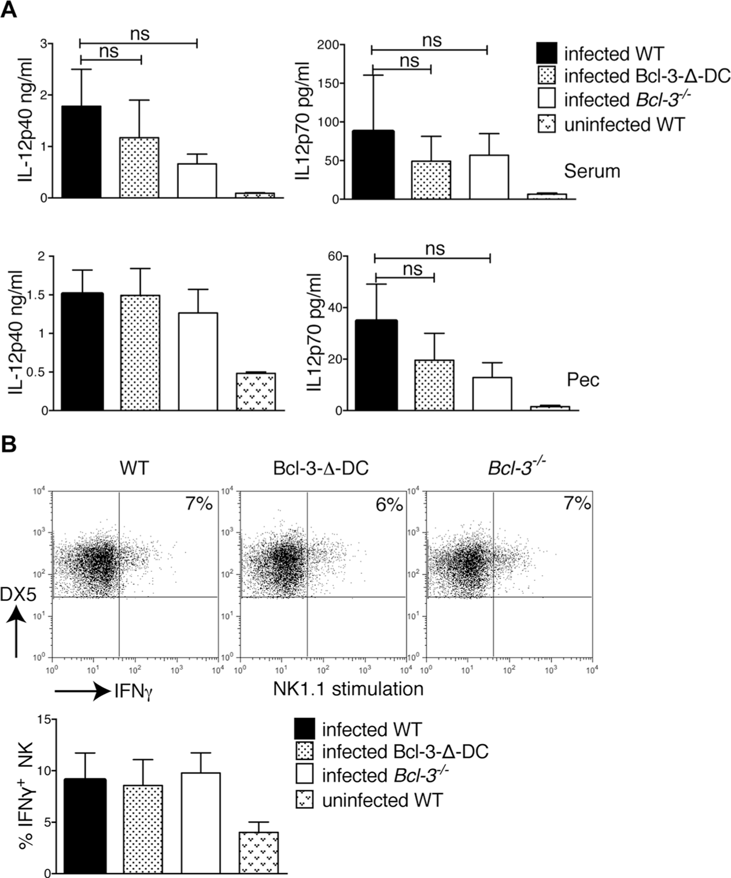
Figure 2.

Innate immune response after T. gondii infection is independent of Bcl-3 expression in CD11c+ cells. (A) WT, Bcl-3−/−, and Bcl-3-∆-DC mice were infected with 20 cysts of T. gondii (ME49 strain). Three days p.i., the levels of IL-12-p40 (left) and IL-12p70 (right) in serum (top) and fluid of peritoneal cavity (PEC, bottom) were assessed by ELISA. Data are shown as mean ± SEM for n = 6 mice/group, pooled from 2 experiments; ns = not significant. (B) Mice were infected as in (A), splenocytes were isolated 3 days later and stimulated with plate-bound NK1.1 for 6 h. Cells were stained for DX5 and CD3, then for intracellular IFN-γ, and IFN-γ production by NK cells was assessed after gating on DX5+CD3− cells. Representative FACS analysis is shown (top) as well as the mean ± SEM; n = 8 mice/group pooled from 2 experiments. Statistical analysis performed by unpaired, two-tailed Student’s t-test.
