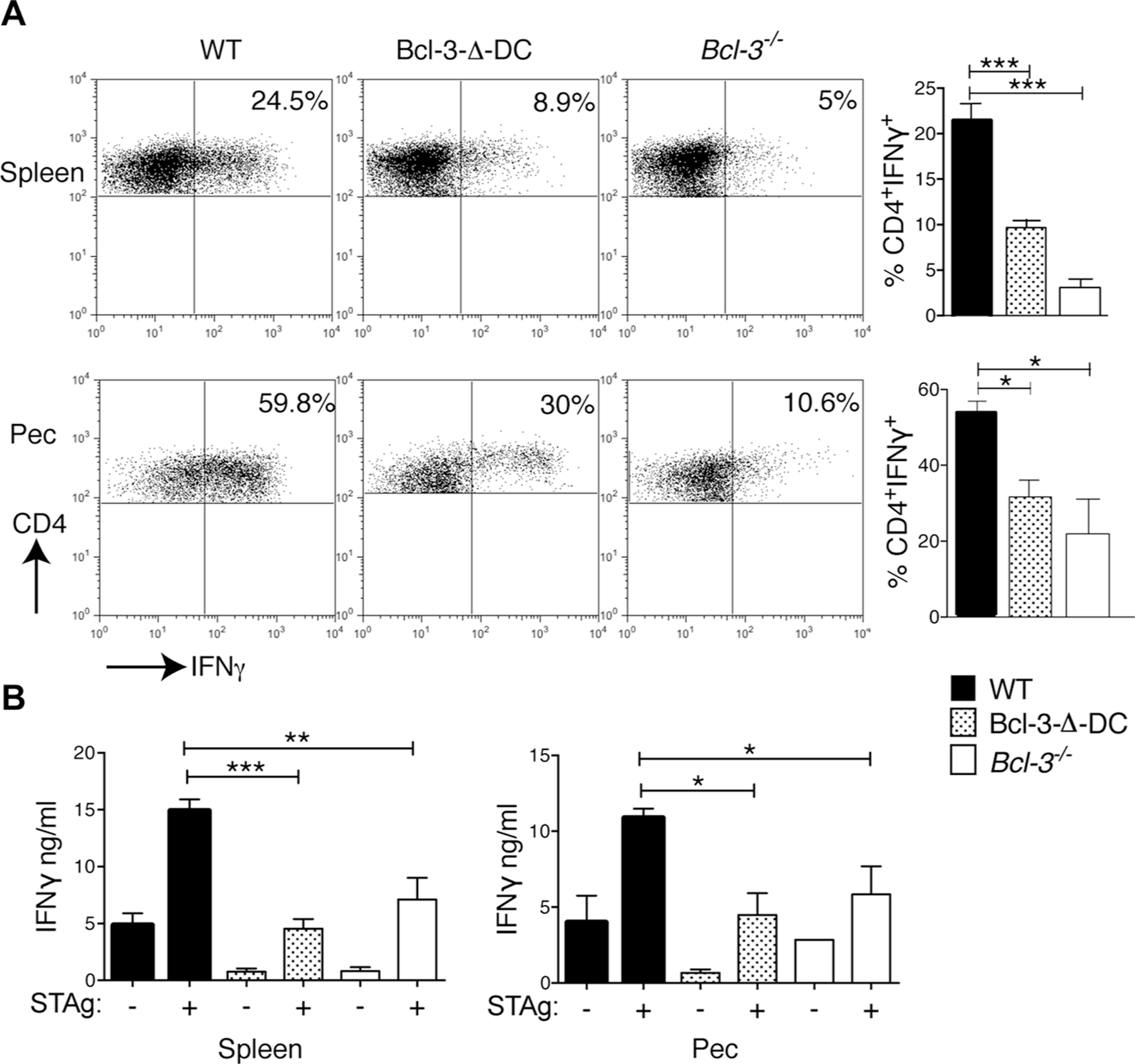
Figure 4.

Bcl-3 expression in DCs is required for CD4-mediated IFN-γ production in response to T. gondii infection. (A) WT, Bcl-3−/−, and Bcl-3-∆-DC mice were infected with T. gondii. Splenocytes (top) and cells from peritoneal cavity (PEC, bottom) were isolated 1 week later, stimulated for 6 h with plate-bound anti-CD3, and intracellular IFN-γ production by CD4+ cells was measured by flow cytometry. Representative plots are shown (left) and data are summarized as mean ± SEM; n = 8 mice/group pooled from 2 experiments (right). (B) CD4+ T-cells isolated from splenocytes (left) and PEC cells (right) of infected mice as in (A) were stimulated in the presence STAg and irradiated WT splenocytes for 72 h and IFN-γ measured with CBA. Data are shown as mean ± SEM; n = 8 mice/group pooled from 2 experiments. *p<0.05, **p<0.01, ***p<0.0001, unpaired, two-tailed Student’s t-test.
