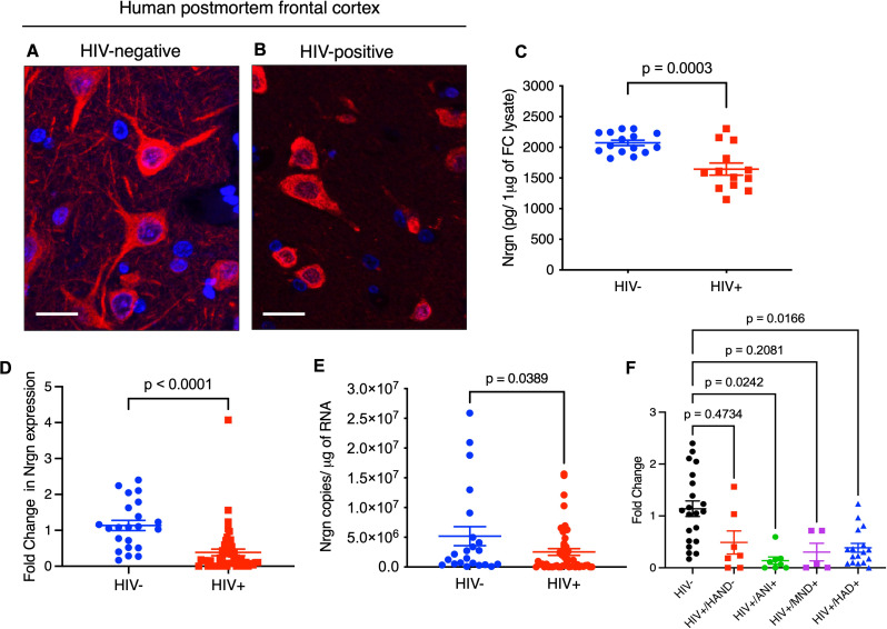
Fig. 1.
Neurogranin (Nrgn) is dysregulated in PWH at the mRNA and protein levels. Representative images of postmortem frontal cortex (FC) neurons stained for Nrgn (Cy3, red) and DAPI (blue) in (A) control and (B) people with HIV-1. The images are z-projections of image stacks acquired at ×60 magnification; scale bar is 10 μm. (C) Quantification of Nrgn levels in whole frontal cortex lysates from people with HIV-1 (N = 13) in comparison with control individuals (N = 15). (D) Relative expression of Nrgn mRNA in FC brain samples from HIV-1 positive individuals (N = 49) compared to control individuals (N = 22) as assessed by RT-qPCR. (E) Quantification of Nrgn mRNA copy number in FC samples of HIV-1 positive individuals compared to control individuals. (F) Comparison of Nrgn expression across different clinical stages of HAND

