
FIGURE 1.
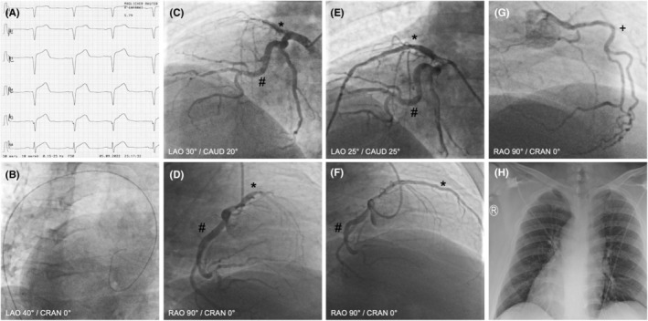
(A) Electrocardiogram showing right precordial leads with ST‐segment elevation in V2R to V5R. (B) Positioning of the guidewire showed the aortic arch bending towards the right side, suggesting organ inversion at LAO 40°/CRAN 0°. (C) Coronary angiography showing proximal thrombotic occlusion of a dominant RIVA at LAO 30°/CAUD 20°. (D) Coronary angiography showing proximal thrombotic occlusion of a dominant RIVA at RAO 90°/CRAN 0°. (E) Angiography after PTCA/stenting at LAO 25°/CAUD 25°. (F) Angiography after PTCA/stenting at RAO 90°/CRAN 0°. (G) Coronary angiography showing left‐sided RCA at RAO 90°/RAN 0. (H) Chest X‐ray showing situs inversus with dextrocardia. *Ramus interventricularis anterior (RIVA), #ramus circumflexus (RCx), +right coronary artery (RCA).