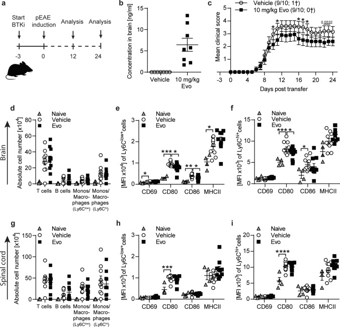
Fig. 3.
In passive EAE evobrutinib downregulates antigen presenting capacity of CNS monocytes/macrophages. C57BL/6 J mice were treated daily with 10 mg/kg evobrutinib or vehicle control started 3 days prior to passive EAE induction with pathogenic T cells. a Overview of experimental setup. b Evobrutinib concentration in brain homogenates isolated from mice 24 days post T cell transfer (n = 7–8). Tissue was collected 30 min after final dose c Group EAE score (n = 9–10). d, g Composition of brain and spinal cord -infiltrating cells (T cells: CD3+, monos/macrophages: CD11b+CD45hiLy6Clow, monos/macrophages: CD11b+CD45hiLy6Chi) were analyzed by flow cytometry on peak of disease (day 12), (n = 8–9). Data are shown as mean fluorescence intensity, (MFI, n = 9–10). e–f, h–i Ly6Chi+ and Ly6C.low+ macrophages/monocytes were isolated from brain (e–f) and spinal cord (h–i) on peak of disease (day 12) and changes in markers involved in activation were analyzed by flow cytometry and are shown as mean fluorescence intensity, (MFI, n = 9–10). The mean ± standard error of the mean is indicated in all graphs. Data sets are representative from at least two independent experiments. Asterisks indicate significant differences calculated using c) unpaired two-tailed t-test (*P ≤ 0.05, **P ≤ 0.01, ***P ≤ 0.001, ****P ≤ 0.0001), d–i one-way analysis of variance corrected by Holm-Sidak (*P ≤ 0.05, **P ≤ 0.01, ***P ≤ 0.001, ****P ≤ 0.0001)

