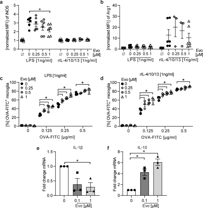
Fig. 6.
BTK inhibition promotes anti-inflammatory microglia as well as human monocyte phenotype and enhances phagocytosis independent of the stimulatory milieu. a, b Primary microglia incubated with indicated evobrutinib concentrations followed by culture in the presence of 1 ng/ml LPS or a mix of anti-inflammatory cytokines (recombinant (r) IL-4/10/13) for 18 h. Expression of iNOS and Arginase 1 (Arg1) were analyzed by flow cytometry and normalized to vehicle control and are shown as mean fluorescence intensity, (MFI, n = 10, pooled from at least 3 independent experiments). c, d Following pre-incubation with indicating concentrations of evobrutinib or vehicle and stimulation with LPS or rIL4/10/13, microglia were cultured in the presence of FITC-labelled ovalbumin (OVA-FITC) for 2.5 h and the frequency of phagocytosing OVA-FITC+ cells (n = 3 wells/condition) was analyzed via flow cytometry. e, f Primary human monocytes were stimulated with 100 ng/mL GM-CSF and treated with evobrutinib or vehicle for 48 h. Gene expression was measured subsequently by qPCR. The mean ± standard error of the mean is indicated in all graphs. If not mentioned, data sets are representative of at least 2–3 independent experiments. Asterisks indicate significant differences calculated using a–d one-way analysis of variance corrected by Holm-Sidak. e, f Student t-test *p < 0.05, compared with GM-CSF control, g, h Kruskal–Wallis non-parametric test with Dunn ‘s multiple comparison post-test compared to medium control. *P ≤ 0.05, **P ≤ 0.01, ***P ≤ 0.001, ****P ≤ 0.0001

