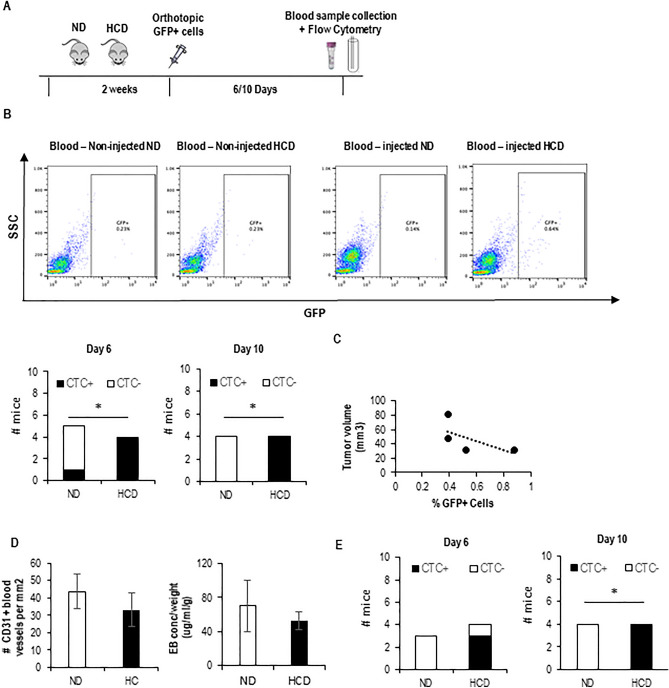
Figure 1.
Short-term HCD potentiates the intravasation of BC cells. (A) Experimental design: mice were fed with either a normal diet (ND) or high cholesterol diet (HCD) for 2 weeks and then injected with GFP-expressing breast tumor cells at the mammary fat pad. Six and ten days after injection, blood samples were collected and analyzed by flow cytometry (FC) to search for the presence of GFP+ cells. (B) Representative plots for the FC analysis of blood samples from mice (BALB/c) that have not been injected with tumor cells (4T1): blood-non-injected (blank), and from blood samples from one representative mouse from each group that have been injected with tumor cells (injected ND and HCD). The bottom graphs show the number of mice in which CTCs were detected 6 days (n = 5 for ND and n = 4 for HCD) and 10 days (n = 4 for both groups) after injecting the tumor cells for each group. To statistically test for a relationship between the presence of CTCs in mice and the diet, a contingency table was used and a Fisher’s exact test applied (*p < 0.05). (C) On day 10, mice were sacrificed, tumors collected, and tumor size determined and plotted against the number of GFP+ tumor cells in circulation for each mouse. The graph shows data from HCD-fed mice, as only these mice had CTCs at day 10. (D) Number of CD31-expressing blood vessels (left) and permeability of blood vessels (right) were quantified for each mouse. Graphs shows means ± SD. (E) Same as in (A) and (B), but for the MDA-231 cell line on NSG mice (n = 3 ND, n = 4 HCD).

