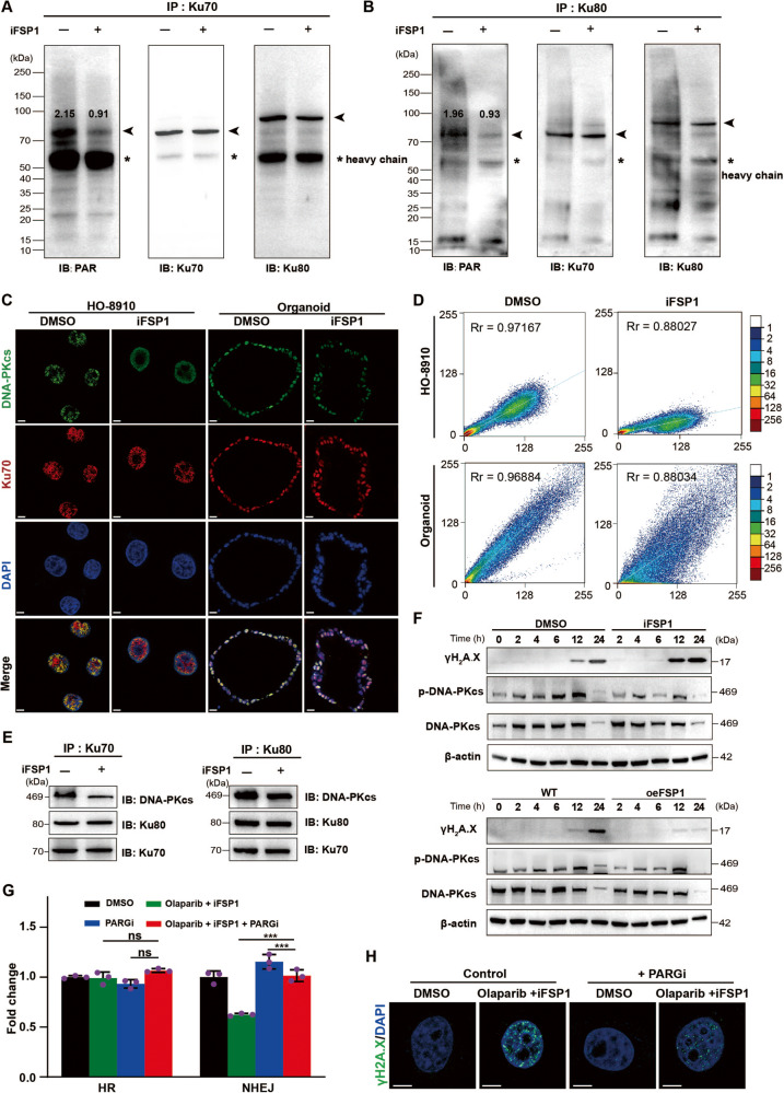
Fig. 6. FSP1 regulates NHEJ activity through PARylation of Ku70.
A, B Effect of FSP1 on PARylation of Ku70 was determined by IP with anti-Ku70 antibody (A) or anti-Ku80 antibody (B) and immunoblots analysis in HEK293t cells. The scaled quantification of PARylation were shown. *: heavy chain. C‒E Immunofluorescence (C) and immunoblots analysis (E) determined ternary complex formation of DNA-PKcs with Ku70/Ku80 complex after FSP1 inhibition in the HO-8910 cells or PDOs. The Pearson’s correlation coefficient of colocalization of Ku70 and DNA-PKcs was calculated (D). Scale bar: 2 μm and 20 μm, respectively. F The immunoblots analysis determined the levels of γH2A.X, DNA-PKcs and phosphate-DNA-PKcs (p-DNA-PKcs) in the HO-8910 cells to the treatment of FSP1 inhibition or overexpressing FSP1. G, H The PARG inhibitor PDD00017273 (PARGi) was used to avoid the PARylation degradation in the HO-8910 cells. The HR and NHEJ activity were determined by the BLRR system (G). ***P < 0.001; ns no significance. The immunofluorescence showed the levels of γH2A.X in each group (H). Scale bar: 5 μm.

