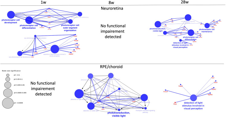
Figure 4.
Decreased functions of neuroretina and RPE/choroid. Functions of neuroretina and RPE/choroid versus time after the start of infection. Examples of significantly perturbed and decreased functions of the retina and RPE/choroid, analyzed in Cytoscape. Large blue nodes with blue text reveals decreased functions. Node size indicates significance as given by the size of the gray nodes. Proteins that participate in the functions are given with red names. In neuroretina, a strong inhibition of neuroretinal function was seen early at 1 week (1 w) of infection with improvement at 8 weeks (8 w) where no functional impairment was detected. Decreased function was also observed late at 28 weeks (28 w) of infection. In the RPE/choroid, no early decreased retinal function was observed. Decreased functions were observed at 8 weeks and at 28 weeks.

