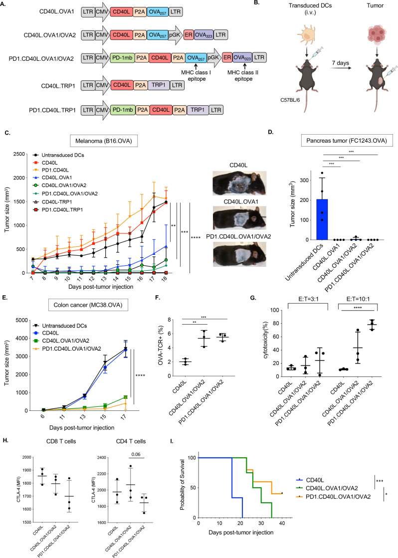
Figure 1.
Immunization with lentiviral vector-transduced DC vaccine vectors slows the growth of solid tumors. (A) The structure of lentiviral vaccine vectors is diagrammed. The vectors express CD40L, OVA class I and class II restricted T cell epitopes OVA257-264 (OVA1) and OVA323-339 (OVA2), and TRP1 and the PD-1 microbody (PD-1mb). (B) DCs were transduced with each lentivirus and then injected intravenously into C57BL/6 mice (1×10⁶ cells). One week later, the mice were inoculated subcutaneously with 2.5×10⁵ B16.OVA cells. (C) The tumor size was measured over 18 days (n=4). (D) DCs were transduced with vaccine vectors and injected intravenously. One week later, the mice were inoculated orthotopically with 1×10⁶ FC1242.OVA tumor cells. Tumor size was measured at 21 days post-DC injection (n=4). (E) Transduced DCs were injected intravenously. After 7 days, the mice were inoculated with 1×10⁶ MC38.OVA colon carcinoma cells. Tumor sizes were measured over 17 days (n=3). (F) Mice were immunized with transduced DCs. After 7 days, the splenocytes were analyzed by flow cytometry with OVA-specific class I tetramers CD8+T cells, gating on CD8+cells. (G) The cytolytic activity of splenocytes from DC-immunized mice against MC38.OVA target cells was measured in an in vitro cytolysis assay. The MC38.OVA target cells were stained with CFSE and incubated for 24 hours with splenocyte effectors at ratios of 1:3 and 1:10. The percentage of lysed cells was then determined by staining with viability dye and analysis by flow cytometry. (H) Survival of the mice following immunization and MC38.OVA implantation is shown (n=5). **p≤0.01, ***p≤0.001, ****p≤0.0001. DCs, dendritic cells ; CD40L, CD40 ligand; OVA, ovalbumin; TRP1, tyrosinase-related protein 1; PD-1, programmed cell death 1; CFSE, carboxyfluorescein succinimidyl ester.

