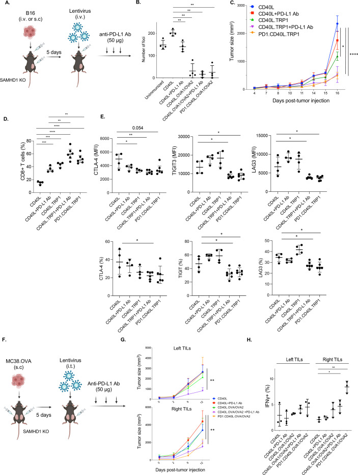
Figure 5.
Lentiviral vaccine and checkpoint inhibitor combination therapy boosts CTL activity. (A) As diagrammed, SAMHD1 KO mice were injected intravenously or subcutaneously with B16.OVA melanoma cells (n=4–6) and, 5 days later, injected intravenously with 3×10⁶ I.U. of lentiviral vectors encoding CD40L, CD40L.TRP1, or PD-1.CD40L.TRP1. In additional groups, the vaccinated mice with CD40L and CD40L.TRP1 were treated three times every other day with anti-PD-L1 antibody starting 5 days postvaccination. (B) The effect of vaccination on the number of metastatic lung foci was determined. SAMHD1 KO mice were injected with B16.OVA intravenously (n=4) and, after 4 days, injected intravenously with lentiviral vaccine vector. An additional set of CD40L and CD40L/OVA1/OVA2 vector-vaccinated mice were treated with anti-PD-L1 antibody (50 µg) injected every 3 days. After 25 days, metastatic foci in the lungs were counted. (C) Tumor size in tumor-bearing mice (subcutaneous injection) was measured beginning on the day of vaccination. (D) The fraction of CD8 T cells in the spleen was determined by flow cytometry (n=4–6). (E) Mean fluorescence intensity (MFI: top) and the percentage (bottom) of exhaustion markers CTLA-4, TIGIT3, and LAG3 on the CD8+T cells were analyzed by flow cytometry. (F) As diagrammed, mice were injected subcutaneously with MC38.OVA on the left and right flanks. The tumor on the right side was then injected with the vaccine lentiviral vector followed by three injections of anti-PD-L1 (n=3). (G) Tumor size on the right and left sides was measured over 12 days starting from the day of vaccination. (H) The number of IFNγ+ CD8+ T cells was analyzed by flow cytometry. *p≤0.05, **p≤0.01, ***p≤0.001, ****p≤0.0001. KO, knockout; IFNγ, interferon gamma; CTL, cytotoxic T lymphocytes; OVA, ovalbumin; PD-L1, programmed death-ligand 1; TRP1, tyrosinase-related protein 1; CTLA4, cytotoxic T-lymphocyte-associated protein 4; TIGIT3, T-cell immunoreceptor with immunoglobulin and immunoreceptor tyrosine-based inhibitory motif domains 3; LAG3, lymphocyte activation gene 3; I.U., infectious unit.

