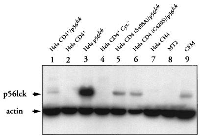
FIG. 2.
Detection of p56lck expression by Western blot analysis. HeLa CD4+/p56lck, HeLa CD4+, HeLa p56lck, HeLa CD4+ Cyt−, HeLa CD4 (C420S)/p56lck, HeLa CD4 (S408A)/p56lck, and HeLa CH4 extracts containing 50 μg of total cellular proteins were electrophoresed in an SDS–10% polyacrylamide gel and blotted to a PVDF membrane. The membrane was incubated with a mixture of anti-p56lck and antiactin MAbs and then reacted with GAM Ig-peroxidase conjugate. Bound MAbs were revealed by incubation of the membrane with ECL reagent and exposure to Hyperfilm-ECL. Controls consist of lysates from MT2 cells (a human T-cell leukemia virus type 1-transformed CD4+ T-cell line which lacks p56lck expression) and CEM cells (a CD4+ T-cell line which expresses p56lck).

