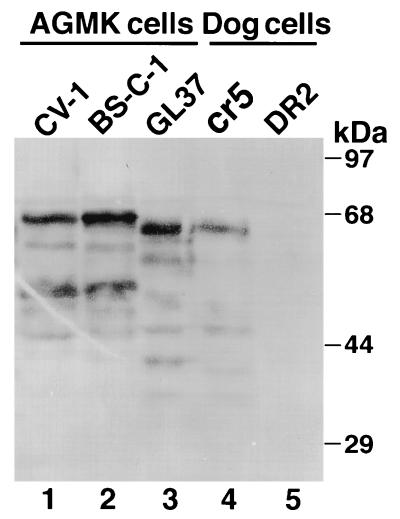
FIG. 2.
Western blot analysis of cytoplasmic extracts of AGMK cell lines. Cytoplasmic extracts of AGMK CV-1 (lane 1), BS-C-1 (lane 2), and GL37 (lane 3) cells and control dog cells transfected with GL37 HAVcr-1 cDNA (cr5 cells [lane 4]) and vector alone (DR2 cells [lane 5]) were prepared in RSB–1% Nonidet P-40. Cytoplasmic extracts were separated by sodium dodecyl sulfate-polyacrylamide gel electrophoresis (10% gel), transferred to nylon membranes, and probed with rabbit anti-GST2 Ab. The positions and sizes of prestained molecular mass markers are shown on the right.

