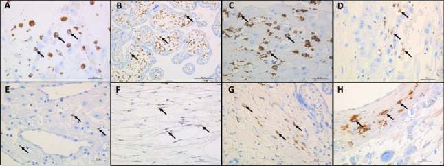
Fig. 2.

Immunohistochemistry of immune cell markers and the SARS-CoV-2 antigen in the placenta of a patient with fetal demise. A Macrophages detected at the interphase between the decidua of membranes and chorion (arrow); anti-CD68 immunohistochemical stain, 40 × magnification, scale bar: 20 µm; B Hyperplasia of macrophages in chorionic villi showed by arrows; anti-CD68 immunohistochemical stain, 20 × magnification, scale bar: 50 µm; C, Macrophages detected in the decidua represented by arrows; anti-CD68 immunohistochemical stain, 40 × magnification, scale bar: 20 µm; D, Natural killer cells in decidua basalis showed by arrows; anti-CD56 immunohistochemical stain, 40 × magnification, scale bar: 20 µm; E CD8 T cytotoxic lymphocytes detected in the decidua basalis represented by arrows; anti-CD8 immunohistochemical stain, 40 × magnification, scale bar: 20 µm; F SARS-COV2-infected stromal cells of the chorion represented by arrows; anti-SARS-CoV-2 nucleocapsid immunohistochemical stain, 40 × magnification, scale bar: 20 µm; G, SARS-CoV-2-infected stromal cells of chorionic villi represented by arrows; anti-SARS-CoV-2 nucleocapsid immunohistochemical stain, 40 × magnification, scale bar: 20 µm; H SARS-CoV-2-infected decidual cells represented by arrows; anti-SARS-CoV-2 nucleocapsid immunohistochemical stain, 40 × magnification, scale bar: 20 µm. Abbreviations: SARS-CoV-2: severe acute respiratory syndrome coronavirus 2
