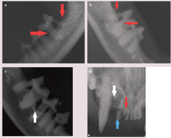
Figure 22.
Alveolar bone loss is evidenced by radiolucency in the coronal area of the bone. Horizontal bone loss (a,b) appears as generalized bone loss of a similar level across all or part of an arcade (red arrows). Vertical (angular) bone loss (c,d) has the radiographic appearance of one area of recession below the surrounding bone (white arrows). Note also in (d) the fractured third incisor (103; blue arrow) and retained root tip of the second incisor (102; red arrow)

