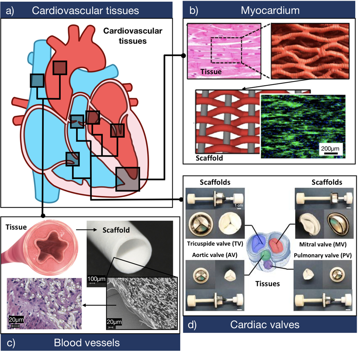
Fig. 2. Biomimetic design criteria for the development of scaffolds for cardiovascular tissue engineering.

a) Schematic of a sagittal plane of the human heart showing the three main tissues that comprise the cardiovascular system. b) Biomaterials used to develop myocardial tissues should provide a highly biocompatible and electroconductive matrix that supports CM function. The panel shows a representative F-actin (green)/DAPI (blue) fluorescent image of CMs growing on a biologically inspired interwoven scaffold. The scaffold was comprised of aligned conductive nanofiber yarns, synthesized from PCL, silk fibroin, and CNTs (Adapted from Ref. [85]). c) Biomaterials used to develop TEBVs should provide adequate burst pressure and mechanical compliance, while also allowing the attachment and proliferation of endothelial cells. The panel shows the histological assessment of endothelial cells growing on a tubular scaffold. The scaffold was comprised of electrospun poly (L-lactide) microfibers (Adapted from Ref. [86]). d) Biomaterials used to develop TEHVs should be able to withstand high trans-valvular pressures while also maintaining low flexural stiffness. The panel shows four types of cardiac valve scaffolds synthesized through double component deposition (DCD) of poly (ester urethane) urea (PEUU) microfibers (Adapted from Ref. [87]).
