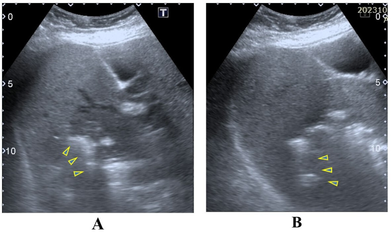
Figure 1 . POCUS image A of gas-forming pyogenic liver abscess (GFPLA) demonstrates an ill-defined margin solitary lesion with heterogenous echogenicity. POCUS image B demonstrates brightly echogenic reflectors with posterior reverberation artefacts noted within the lesion. Yellow triangles on both images show the posterior reverberation artefacts.

