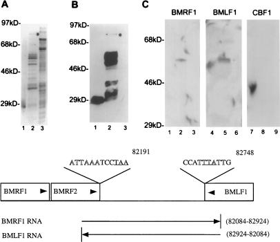
FIG. 5.
Mta binds target mRNAs. Purified GST-Mta, control GST, and crude control MBP-Tax were separated by SDS-PAGE and transferred to Immobilon-P filters. The membrane-bound proteins were denatured and renatured as described in the text and incubated with the indicated RNA probes. (A) Silver stain of purified GST (lane 1), purified GST-Mta (lane 2), and crude MBP-Tax (lane 3). The full-length GST-Mta is indicated by a dot. The gel shows numerous background proteins that are available for nonspecific probe binding. (B) A membrane blot containing the proteins shown in panel A was immunoprobed with anti-GST MAb and visualized by chemiluminescence. The purified GST-Mta was partially degraded. Note that the degradation was largely progressive from the carboxyl terminus since most degradation products tested positive for the amino-terminal GST fusion partner (lane 2). (C) A membrane blot of the proteins shown in panel A was incubated with BMRF1 and BMLF1 RNA probes and a control probe transcribed from the cellular CBF1 cDNA. GST-Mta, but not GST or MBP-Tax, bound to the BMRF1 and BMLF1 RNA probes (compare lanes 2 and 5 to lanes 1, 3, 4, and 6). The control CBF1 RNA did not bind to GST-Mta (lane 8). (D) Map locations of the BMRF1 and BMLF1 probes relative to the BMRF1, BMRF2, and BMLF1 ORFs. The map coordinates are those for the B95-8 genome (4). Arrowheads indicate the direction of transcription of the genes and probe RNAs. The polyadenylation signals for these genes are shown in outlined type, and the translation termination codons for BMRF2 and BMLF1 are underlined.

