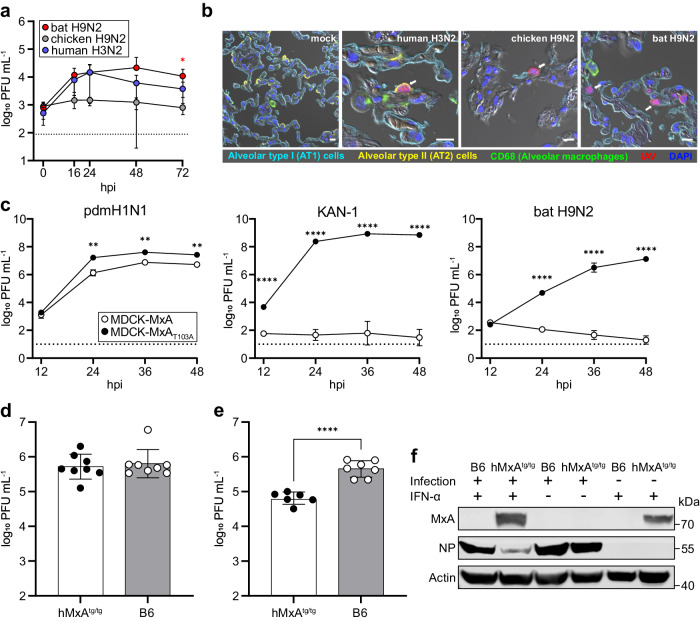
Fig. 2. Bat H9N2 replicates in human lung explants and suppresses induction of MxA in MxA-transgenic mice.
a Human lung tissue explants (n = 4) were infected with human H3N2, chicken H9N2 or bat H9N2 with 1 × 10⁶ PFU, and viral titers were determined at the indicated time points. Error bars indicate standard deviation and statistical analysis was performed using non-paired, non-parametric Kruskal-Wallis test (*p = 0.0324). Data are mean ± SD of n = 4 independent experiments. Dashed line indicates detection limit (b) At 24 hpi, human lung explants were stained for alveolar type I (AT1) (cyan) and type II (AT2) cells (yellow), CD68 indicating alveolar macrophages (green) and IAV antigens (red). Note, in chicken H9N2 and bat H9N2 infected cells, AT2 labeling was omitted for better visualization. White arrows indicate infected cells. Scale bar, 10 µm. c MDCK cells overexpressing MxA or inactive MxAT103A were infected with human-adapted pdmH1N1, avian KAN-1 (H5N1) or bat H9N2 at an MOI of 0.001, and viral titers were determined at the indicated time points. Data are mean ± SD of n = 3 independent experiments; statistical analysis was performed using two-tailed t-tests; **P = 0.01; ****P = 0.0001. Dashed line indicates detection limit. d hMxAtg/tg (n = 8) or wild-type B6 mice (n = 8) were infected with 1 × 10⁴ PFU. Lung viral titers were determined 3 dpi. e hMxAtg/tg (n = 6) or wild-type B6 mice (n = 7) were pretreated with IFN-α 18 h prior to infection with 1 × 10⁴ PFU. Lung viral titers were determined 3 dpi. Data are mean ± SD; statistical analysis was performed using two-tailed t-tests; ****P = 0.0001. f MxA, NP and actin protein levels in homogenized lungs from IFN-α pretreated or infected mice from (d,e) were detected by Western blot. Source data are provided as a Source Data file.

