Figure 3.
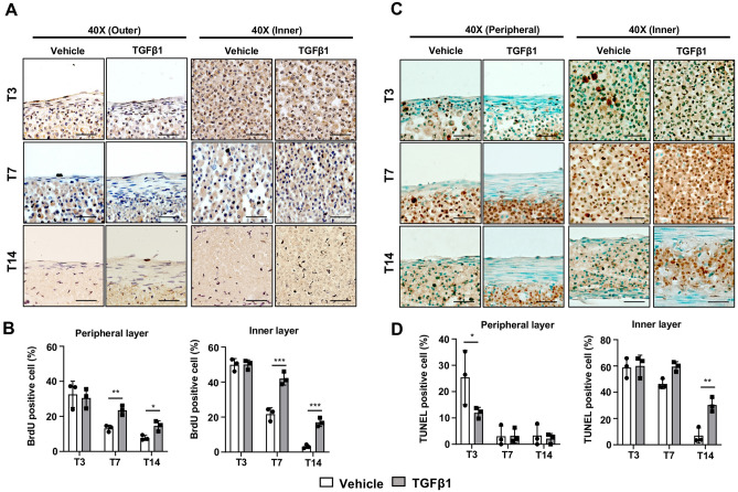
Cellular proliferation and apoptosis in the 3D tendon constructs. (A) BrdU staining of peripheral and inner layers in vehicle- and TGFβ1-treated 3D tendon constructs at each stage (T3, T7, and T14). Scale bars indicate 50 μm. (B) Quantification results of BrdU-positive cells (brown) in peripheral and inner layers of 3D tendon constructs (** indicates P < 0.01 and *** indicates P < 0.001, n = 3). (C) TUNEL assay of peripheral and inner layers in vehicle- and TGFβ1-treated 3D tendon constructs at each stage (T3, T7, and T14). Scale bars indicate 50 μm. (D) Quantification results of TUNEL-positive cells (brown) in peripheral and inner layers of 3D tendon constructs (* indicates P < 0.05 and ** indicates P < 0.01, n = 3 constructs per group).

