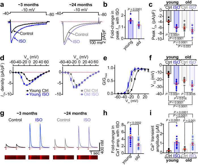
Fig. 1. β-AR-stimulated augmentation of ICa and Ca2+ transients is diminished in aging.
a Representative whole-cell currents elicited from young and old ventricular myocytes before (control; black) and during application of ISO (blue). b Dot-plots showing the fold-change in peak current with ISO in young and old myocytes. c Dot-plots showing peak ICa density before and after ISO. d Plots showing the voltage dependence of ICa density for both groups before and after ISO. e Voltage dependence of the normalized conductance (G/Gmax) fit with Boltzmann functions and f dot-plots showing the V1/2 of activation for each group. N-numbers for patch clamp data in (b–f ) is as follows: young (N = 9, n = 13) and old (N = 5, n = 11). g Representative Ca2+ transients recorded before (black) and after ISO (blue) from paced young and old myocytes. h Dot-plots showing the fold increase in Ca2+ transient amplitude after ISO and i Ca2+ transient amplitude before and after ISO. N-numbers for Ca2+ transient data in (h, i) is as follows: young (N = 5, n = 15) and old (N = 3, n = 15). Unpaired two-tailed Student’s t-tests were performed on data sets displayed in (b, h). Two-way ANOVAs with multiple comparison post-hoc tests were performed on data displayed in (c, f, i). Young data in (b–f ) is pooled from NIA young and JAX young myocytes, as no significant differences were found in ICa when NIA young and JAX young myocytes were compared (see Supplementary Fig. 1a–e). Young data in (h, i) is from NIA young. Data are presented as mean ± SEM. Source data are provided in the Source Data file.

