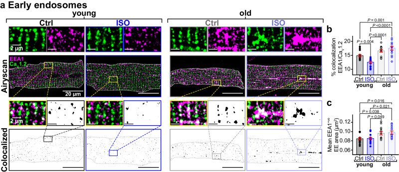
Fig. 4. Aging impairs β-AR-stimulated CaV1.2 recycling.
a Airyscan super-resolution images of CaV1.2 (green) and EEA1 (magenta) immunostained myocytes with and without ISO. Bottom: Binary colocalization maps show pixels in which CaV1.2 and EEA1 completely overlapped. b dot-plots summarizing % colocalization between EEA1 and CaV1.2 young (control: N = 3, n = 16; ISO: N = 3, n = 16) and old (control: N = 3, n = 14; ISO: N = 3, n = 14) myocytes, and c EEA1-positive endosome areas in young (control: N = 3, n = 15; ISO: N = 3, n = 16) and old (control: N = 3, n = 14; ISO: N = 3, n = 13) myocytes. Data were analyzed using two-way ANOVAs with multiple comparison post-hoc tests. Young data in (b, c) are from JAX young. Note no significant differences in EEA1/CaV1.2 colocalization, responsivity to ISO, or endosome size was detected when young JAX and young NIA myocytes were compared (Supplementary Fig. 1h, i). Data are presented as mean ± SEM. Source data are provided in the Source Data file.

