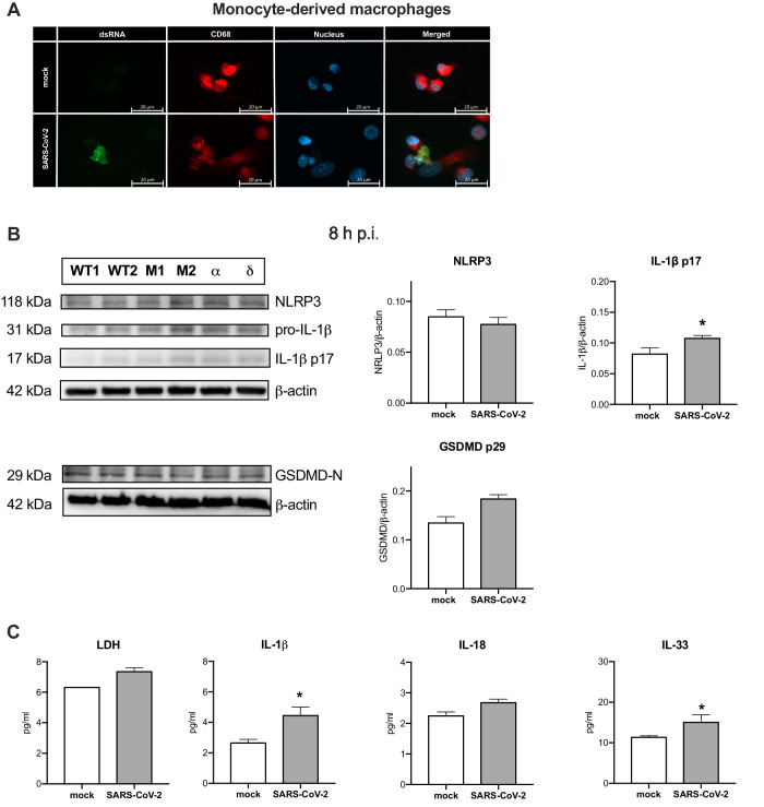
Fig. 6. SARS-CoV-2 infection of monocyte-derived macrophages elicits inflammasome signaling.
Human monocyte-derived macrophages were infected with SARS-CoV-2 at MOI 1 for 8 h. A Confocal microscopy analysis of the presence of SARS-CoV-2 dsRNA in CD68+ macrophages. B Western blot analysis of the expression of NLRP3, IL-1β (pro and cleaved p17), GSDMD-N and β-actin as loading control (left) upon infection with wild type, α and δ SARS-CoV-2 strains, M1, M2 – mock; densitometric analysis (right). C Concentration of LDH, IL-1β, IL-18, and IL-33 was analyzed in the supernatant from the infected macrophages. Graphs represent means ± SEM, n = 3. Two groups comparisons were analyzed with t-test. *p < 0.05.

