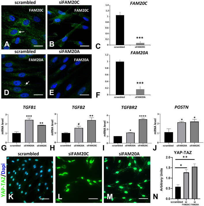
Figure 9.
FAM20C and FAM20A silencing. (A–G) GFs were transfected with siRNAs targeted either FAM20C, FAM20A or scrambled siRNAs used as controls. Three days after transfection, cells were analyzed for FAM20C or FAM20A expression using immunocytochemistry (A,B,D,E) or QPCR (C,F). (A) FAM20C protein was localized to endoplasmic reticulum in GFs transfected with scrambled siRNAs. (B) This staining was not observed in GFs transfected with siRNAs targeted FAM20C. (C) Using real time RT-PCR, a 90% decrease in mRNA to FAM20C was observed. (D) FAM20A protein was localized in large vesicles in GFs transfected with scrambled siRNAs. (E) This staining was absent in GFs transfected with siRNAs targeting FAM20A. (F) Using real time RT-PCR, a significant 80% decrease in mRNA to FAM20A was observed. (G–J) Real Time RT-PCR analysis of candidate genes corresponding to TGFB1 (G), TGFB2 (H), the receptor TGFBR2 (I) and Periostin POSTN (J). Scrambled siRNAs, siFAM20C or siFAM20A values correspond to the mean of 3 independent experiments in triplicates of three GFs cultures. Datas represent mean fold gene expressions ± s.d. Data was analyzed via one-way ANOVA with Bonferroni multiple comparisons test (*p < 0.05, **p < 0.01, ***p < 0.005, ****p < 0.001). (K–M) YAP-TAZ immunocytochemistry showed some lightly immunoreactive nuclei in GFs transfected with scrambled siRNAs (K). In GFs transfected with siFAM20C or siFAM20A a dramatic increase in the staining was observed in all nuclei (L,M). (N) Graph representation of the relative fluorescence intensity of YAP-TAZ in GFs transfected with scrambled siRNAs, siFAM20C or siFAM20A. Statistically significant differences are shown as ***P < 0.001. Scale bars: (A,B,D,E) = 20 µm; (K–M) = 50 µm.

