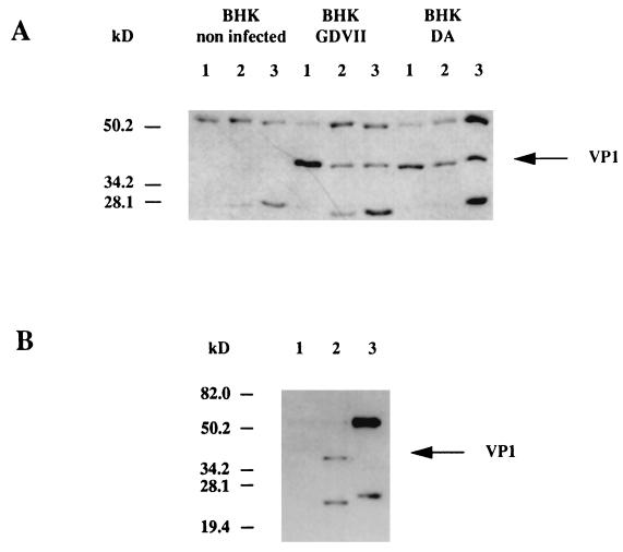
FIG. 5.
(A) Immunoprecipitation of BHK-21 cell lysates. The cells were either not infected or were infected with the GDVII or DA virus. The lysates were immunoprecipitated with the anti-VP1 MAb (lanes 1), the anti-desmin MAb (lanes 2) or the anti-vimentin MAb (lanes 3). Immunoprecipitated proteins were separated by SDS-PAGE (10% polyacrylamide), transferred to a nitrocellulose membrane, and analyzed by Western blotting with the anti-VP1 MAb as described in Materials and Methods. (B) Immunoprecipitation of BHK-21 cell lysates. The cells were either not infected (lane 1) or infected with the GDVII virus (lanes 2 and 3). The lysates were immunoprecipitated with the anti-VP1 MAb (lanes 1 and 2) or an anti-α-actin MAb (lane 3). Immunoprecipitated proteins were separated by SDS-PAGE (10% polyacrylamide), transferred to a nitrocellulose membrane, and analyzed by Western blotting with the anti-VP1 MAb as described in Materials and Methods.

